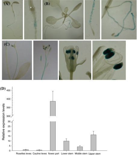
Fig. 3.
Expression pattern of the IGI1 gene. a–c The expression pattern was detected using a GUS reporter gene under control of the IGI1 gene promoter. a–b. Histochemical staining of GUS activity in 5- (a) and 10-day-old (b) seedlings. Expression was detected only in the root hair. c Histochemical staining of GUS activity in each part of a 33-day-old plant. The pictures show the cauline inflorescence and leaf, apical part of main inflorescence, flower, and anther (left to right). GUS expression was weakly detected in the upper stem and immature siliques and strongly detected in the anther of the flowers. d The relative expression levels of the IGI1 gene in different tissues. The flower part has a higher expression level than the other parts. Actin was used for normalization and the error bars indicate the standard deviation

