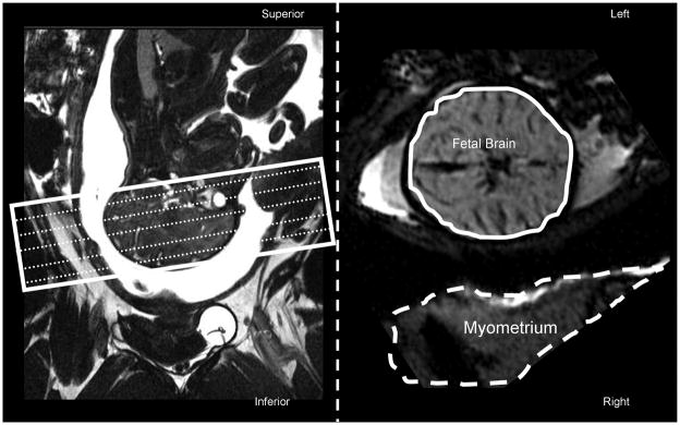
Figure 1.
Imaging slices for dynamic susceptibility imaging (white rectangle) were prescribed axially, parallel to the anterior-posterior commissure plane using a sagittal localizer image (Left panel). Right panel shows the axial slice of the fetal brain at the level of mesencephalon and the dam’s uterine muscle, myometrium. Temporal signal intensity trends were analyzed by manually drawing two volumes of interest: one covered the entire fetal brain (solid contour) and the other covered dam’s myometrium (dashed contour).

