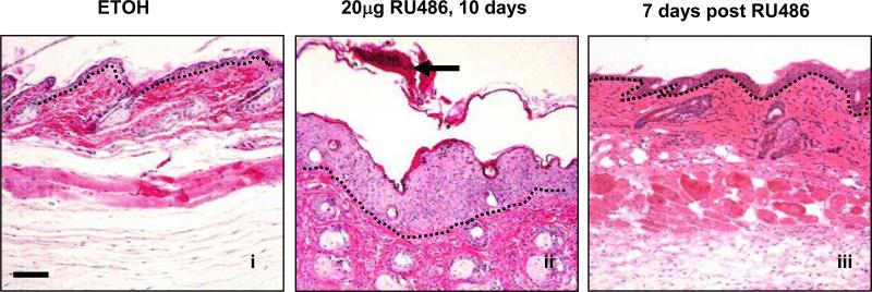
Fig. 1. Effects of TGFβ1wt transgene induction in gene-switch-TGFβ1wt skin.
The H&E staining of dorsal skin from TGFβ1wt gene-switch mice treated with ETOH (i) or RU486(ii) for 10 days. Epidermal hyperplasia and corneal microabscess (arrow) were noticed upon TGFβ1wt induction (ii) and recovered to normal skin after withdrawal of RU486 (iii). The bar in panel (i) represents 100μm for all sections. The dotted line in each section highlights the epidermal-dermal junction.

