FIG. 3.
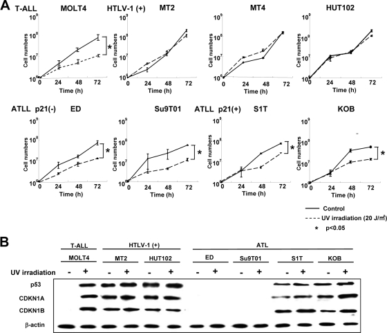
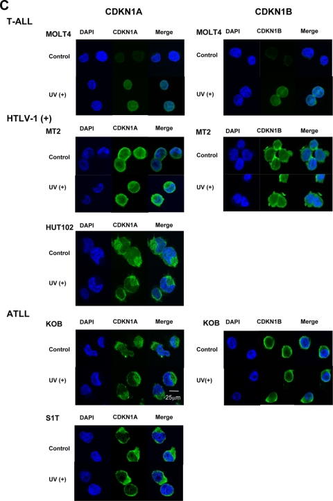
Loss of functional CDKN1A response to UV irradiation in HTLV-1-infected and ATLL cell lines. (A) Cell growth curves of an HTLV-1-negative T-ALL cell line (MOLT4), three HTLV-1-infected cell lines (MT-2, MT-4, and HUT102), and four ATLL-derived cell lines (ED, Su9T01, S1T, and KOB) before and after UV irradiation. Viable cells were counted using the trypan blue exclusion method at the indicated times after 20 J/m2 UV irradiation. Samples at 0 h were nonirradiated controls. A Student's t test was used for statistical analysis. (B) Western blot analyses of p53, CDKN1A, and CDKN1B following UV irradiation. Each cell line, either untreated (−) or UV irradiated (+; 20 J/m2),was cultured for 72 h to examine protein expression. (C) Subcellular localization of CDKN1A and CDKN1B with or without (control) UV irradiation. Localization of CDKN1A and CDKN1B was detected by confocal immunofluorescence analysis. DAPI stain was used to visualize the nuclei. Magnification, ×400; bar, 25 μm.


