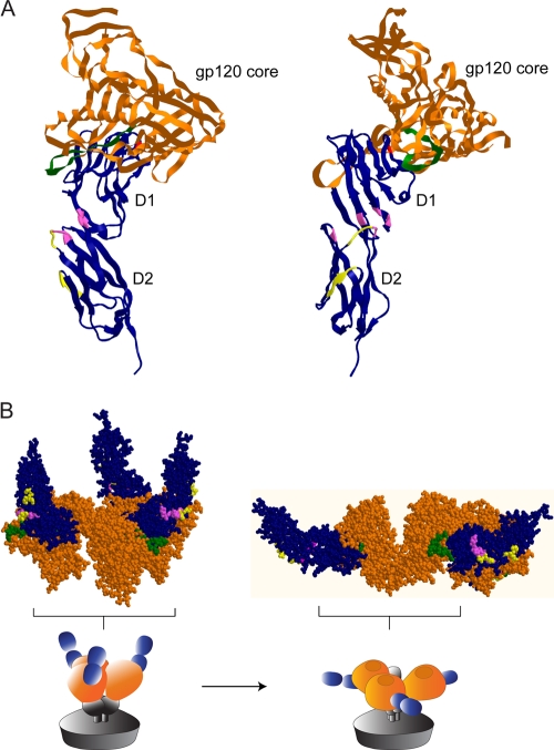
FIG. 4.
Localization of ibalizumab and M-T441 epitopes on three-dimensional structures of hCD4. The epitopes of ibalizumab (pink) and M-T441 (yellow) are highlighted on CD4 (blue) complexed to the core of gp120 (brown), with two views of the structure rotated 90° around a central vertical axis (A), or space-filling model of three CD4s (blue) bound to the trimer of gp120 core (brown), before and after a conformational rearrangement (B), as derived from the cryo-electron microscopy study of Liu et al. (17). The red dot in D1 denotes F43, a critical component that binds HIV-1 Env. The parts in green highlight the V5 loop in gp120.

