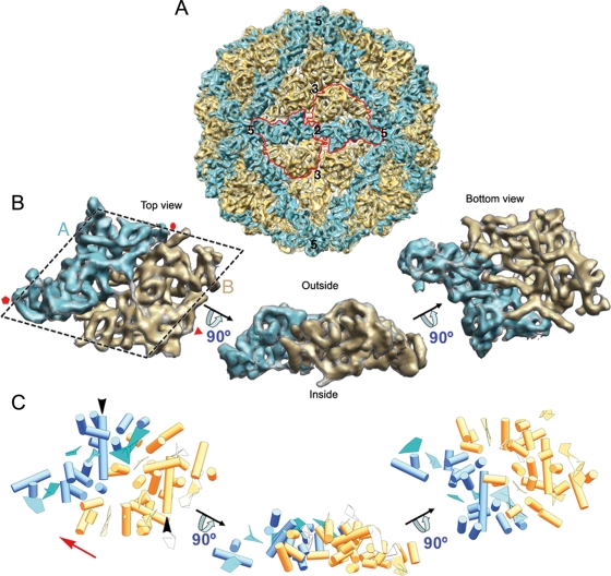
FIG. 3.
Structure of the PcV T=1 capsid and model of the capsid protein fold. (A) Surface-shaded virion capsid viewed along an icosahedral 2-fold axis with capsid protein (CP) elements in cyan and yellow; boundaries for two CPs are outlined in red. Icosahedral symmetry axes are numbered. (B) Segmented asymmetric unit (PcV CP monomer). The dashed line highlights the rhomboidal shape. Protein halves A (cyan) and B (yellow) are indicated. Red symbols indicate icosahedral symmetry axes. (C) PcV CP secondary structural elements (SSE), using the same color scheme and orientations as in panel B. Cylinders, α-helices; planks, β-sheets. The red arrow indicates translation direction to superimpose half-protein B on A. Black arrows indicate the ∼37-Å-long α-helices of both PcV CP elements.

