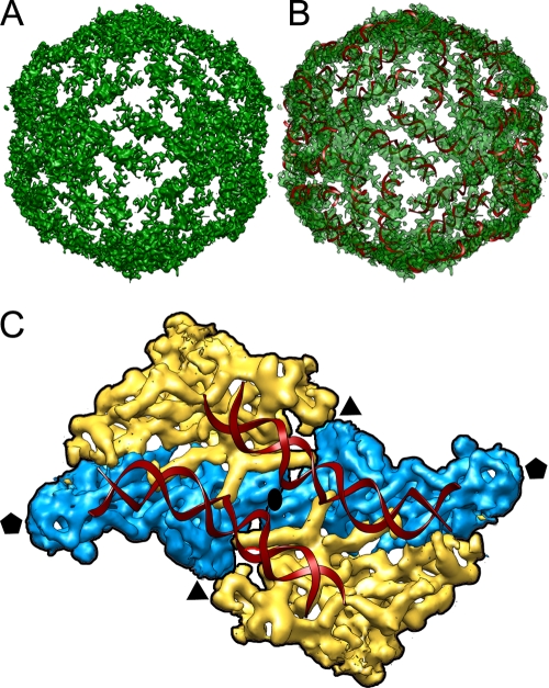
FIG. 7.
Outer layer of dsRNA. (A) Genome density ordered into an icosahedral cage in close contact with the capsid inner surface. The capsid protein has been stripped away, showing the top half of the first layer of ordered dsRNA density. (B) A-form helices of dsRNA docked into the tubular densities corresponding to the dsRNA density. (C) Close-up view down a 2-fold axis from inside, showing two adjacent structural subunits colored as in Fig. 3. The phosphate backbone is traced as a red ribbon for the two dsRNA A-form strands. Icosahedral symmetry axes are indicated.

