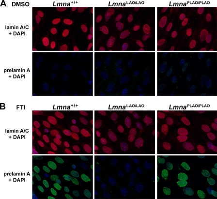
FIGURE 5.
Immunofluorescence microscopy of Lmna+/+, LmnaLAO/LAO, and LmnaPLAO/PLAO fibroblasts with an antibody against lamin A/C (red) and a rat monoclonal antibody against prelamin A (green). These images reveal a higher frequency of misshapen nuclei in LmnaLAO/LAO fibroblasts. Cells were grown in the presence of vehicle (DMSO) alone (panel A), or with an FTI (ABT-100, 2 μm) (panel B). Treatment of cells with an FTI results in an accumulation of prelamin A in Lmna+/+ and LmnaPLAO/PLAO fibroblasts, but not in LmnaLAO/LAO fibroblasts.

