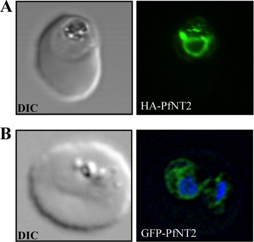
FIGURE 2.
Immunolocalization of HA-PfNT2 and GFP-PfNT2. Anti-HA (A) or anti-GFP (B) antibody (green) localizes to a compartment within transgenic parasites expressing either HA-PfNT2 (A) or GFP-PfNT2 (B). The differential contrast image (DIC) images show the location of the parasites within the erythrocyte. The nucleic acids of the GFP-PfNT2 parasites were visualized using Hoescht 33258 (blue).

