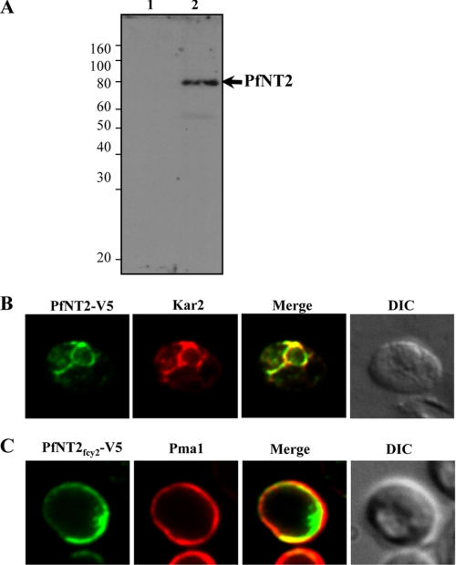
FIGURE 5.
Immunofluorescence analysis of PfNT2 in yeast cells. A, imunoblotting using extracts from yeast cells transformed with either pYES2.1 (lane 1) or pYES2.1-PfNT2CO-V5 (lane 2). Cell lysates were subjected to SDS-PAGE, transferred to nitrocellulose, and probed with an anti-V5 antibody. B and C, immunofluorescence assays in yeast cells transformed with the plasmid encoding V5-tagged PfNT2CO (B) or V5-tagged PfNT2fcy2 (C). Colocalization studies were performed using anti-Kar2 (B) and anti-Pma1 (C) antibodies. Images represent localization of full-length PfNT2 and chimeric PfNT2fcy2 in end3Δ yeast cells. Similar results were obtained in wild type cells (data not shown). DIC, differential contrast image.

