FIGURE 2.
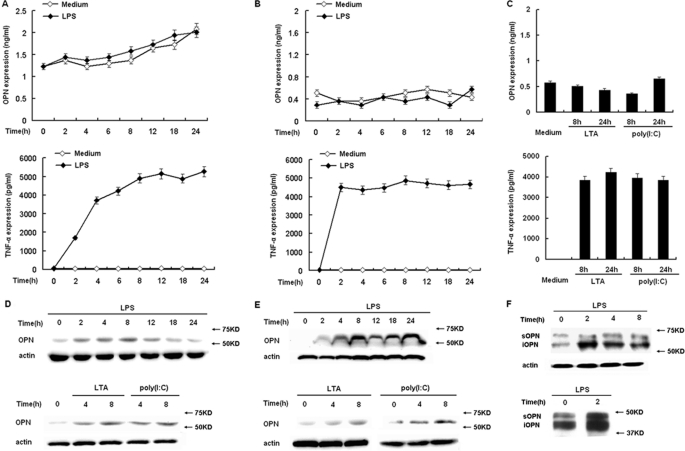
TLRs differentially induce intracellular and secreted OPN expression in macrophages. A, RAW264.7 macrophages or B, peritoneal macrophages were treated with 100 ng/ml LPS as indicated time period. C, peritoneal macrophages were treated with 1 μg/ml LTA or 10 μg/ml poly(I:C) as indicated. Secreted OPN and TNF-α in supernatants were measured by ELISA. D, RAW264.7 macrophages or E, peritoneal macrophages were treated with 100 ng/ml LPS, 1 μg/ml LTA, or 10 μg/ml poly(I:C) as indicated. Intracellular OPN expression was detected by Western blot with R&D OPN Ab. F, peritoneal macrophages were treated with 100 ng/ml LPS as indicated time period. Opn-s and Opn-i were detected by immunoblotting with OPN 2A1 Ab from Santa Cruz Biotechnology (upper panel). OPN from control and LPS-stimulated cells was dephosphorylated and deglycosylated using bovine alkaline phosphatase and peptide N-glycosidase F, respectively, then Opn-s and Opn-i were detected by immunoblotting with OPN 2A1 Ab (lower panel). Actin was used as a cytoplasmic protein loading control. Data are shown as mean ± S.D. (n = 3) of one representative experiment.

