FIGURE 7.
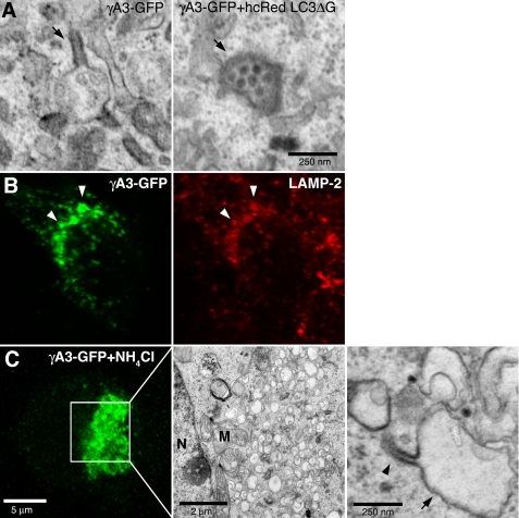
Lysosomes are the substrates for tubulogenesis. A, electron-dense organelles in γA3-GFP and hcRed-LC3ΔG-cotransfected cells resemble lysosomes (right panel, arrow) versus organelles from γA3-GFP-tranfected cells in which the tubules (left panel, arrow) retained electron-dense properties whereas lysosomal-like organelles were less electron-dense. B, colocalization of γA3-GFP with lysosomal marker LAMP-2 indicates Pcdh-γs target to lysosomes, implicating them as the substrate for tubule growth. C, NH4Cl inhibition of lysosomal acidification reduces tubulogenesis. Enlarged vacuoles (arrow) only occasionally sprouted tubules (arrowhead). N, nucleus; M, mitochondria.

