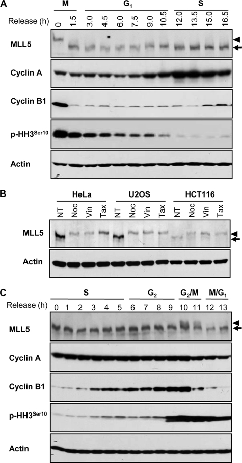
FIGURE 1.
MLL5 displays slower gel mobility at G2/M transition. A, expression of MLL5 in HeLa cells released from the G2/M boundary. B, expression of MLL5 in asynchronous (no treatment (NT)) and G2/M-arrested HeLa, U2OS, and HCT116 cells. G2/M synchronization was achieved by treatment with nocodazole (Noc), vinblastine (Vin), or taxol (Tax). C, expression of MLL5 in HeLa cells that were released from G1/S synchronization. Cyclin A, cyclin B1, and phosphohistone H3Ser10 serve as cell cycle progression markers. Actin serves as a protein loading control. Arrows indicate MLL5, and arrowheads denote the slower migrating form of MLL5 at G2/M phase.

