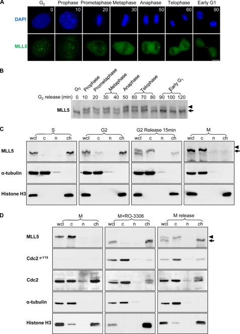
FIGURE 5.
Phospho-MLL5 dissociates from mitotic chromosomes. HeLa cells were grown on coverslips, and G2-arrested cells were released into fresh medium. Samples for immunofluorescence staining and Western blotting were collected every 10 min. A, MLL5 formed intranuclear foci in G2 phase (time 0), but it dissociated from condensed chromosomes and displayed cytosolic staining pattern during mitosis (10 min, prophase; 20 min, prometaphase; 30 min, metaphase; 50 min, anaphase; and 60 min, telophase). When cells completed mitosis (90 min), MLL5 re-localized to the nucleus. Scale bar, 10 μm. DAPI, 4′,6-diamidino-2-phenylindole. B, time course study on the phosphorylation of MLL5 during mitosis. C, HeLa cells were arrested at difference cell cycle stages, and cellular fractionation was performed. Cells arrested in S phase were collected after G1/S release for 4 h; G2 phase arrest was achieved by incubation with RO-3306 for 20 h, and M phase cells were synchronized by the Thy-nocodazole method. α-Tubulin and histone H3 were employed as cytoplasmic (c) and chromatin-associated (ch) protein marker, respectively, and nucleoplasmic protein was denoted as group (n). Whole cell lysate (wcl) serves as total cellular protein control. Phosphorylation and subcellular localization of MLL5 were examined by Western blotting. MLL5 was extracted in the chromatin fraction in S and G2 phase cells (1st and 2nd panel), and in mitotic cells the MLL5 was phosphorylated and extracted in the cytoplasmic fraction (3rd and 4th panels). D, mitotic phosphorylation and localization of MLL5 were dependent on Cdc2 kinase activity. In mitosis-arrested HeLa cells, MLL5 was phosphorylated and extracted in the cytoplasmic fraction (left panel). When mitotic HeLa cells were treated with RO-3306 for 1.5 h, MLL5 was dephosphorylated and extracted in the chromatin fraction. Inhibition of Cdc2 activity was revealed by the increase in phosphorylation of Cdc2 on Tyr-15 (middle panel). When mitotic cells were released into complete medium for 1.5 h and re-entered into the G1 phase, MLL5 was dephosphorylated and associated with chromatin (right panel). Phospho-MLL5 was denoted by an arrowhead.

