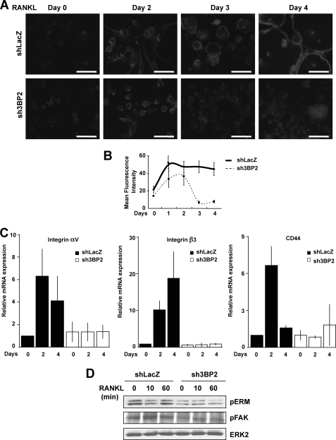
FIGURE 3.
Effects of 3BP2 suppression on RANKL-induced organization of the osteoclast actin cytoskeleton. A, shLacZ and sh3BP2 cells were seeded onto sterile chamber slide treated with sRANKL (40 ng/ml) for the indicated times. After washing, the cells were fixed, permeabilized, and incubated with 100 ng/ml of Texas Red phalloidin for 30 min. Polymerized actin was visualized using a fluorescence microscope. The data shown are representative of three independent experiments. Scale bars, 50 μm. B, cells were treated with sRANKL (40 ng/ml) for the indicated times. Following cell fixation and labeling with AlexaFluor 488-conjugated phalloidin (100 ng/ml), actin polymerization was quantified by flow cytometry. The data are expressed as mean channel fluorescence intensity for each sample. The results represent the means ± S.D. of three independent determinations. C, shLacZ and sh3BP2 cells were stimulated or not with RANKL (40 ng/ml) for the indicated times. The expression of integrin αv and β3 subunits and CD44 mRNA was determined by real time quantitative PCR. The data are expressed as the means ± S.D. of triplicate determinations and are representative of three independent experiments. D, cells were cultured without serum for 12 h before stimulation with sRANKL (100 ng/ml) for 0, 10, and 60 min. The cell lysates were subjected to immunoblot analysis with antibodies against phospho-ERM and phospho-FAK. The same membrane was stripped and reprobed with anti-ERK2.

