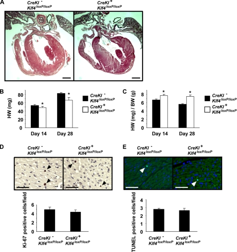
FIGURE 5.
The rates of proliferation and apoptosis were unaltered in the hearts of SM22α-CreKI+/Klf4loxP/loxP mice. A, hearts from SM22α-CreKI−/Klf4loxP/loxP and SM22α-CreKI+/Klf4loxP/loxP mice at P28 were sectioned and stained with Russell-Movat pentachrome method. Representative pictures are shown (n = 6 per each genotype). Bars, 1 mm. B and C, heart weight and the ratio of heart to body weight were measured in SM22α-CreKI−/Klf4loxP/loxP and SM22α-CreKI+/Klf4loxP/loxP mice at P14 and P28. *, p < 0.05 compared with SM22α-CreKI−/Klf4loxP/loxP mice. D and E, Ki-67 staining (D) and TUNEL staining (E) were performed in the heart of SM22α-CreKI−/Klf4loxP/loxP and SM22α-CreKI+/Klf4loxP/loxP mice at P28 (n = 5 per each genotype). Ki-67 staining was visualized by DAB, and sections were counterstained with hematoxylin. TUNEL staining was visualized by fluorescein isothiocyanate, and sections were counterstained with 4′,6-diamidino-2-phenylindole. Arrowheads indicate stained cells. Bars, 50 μm.

