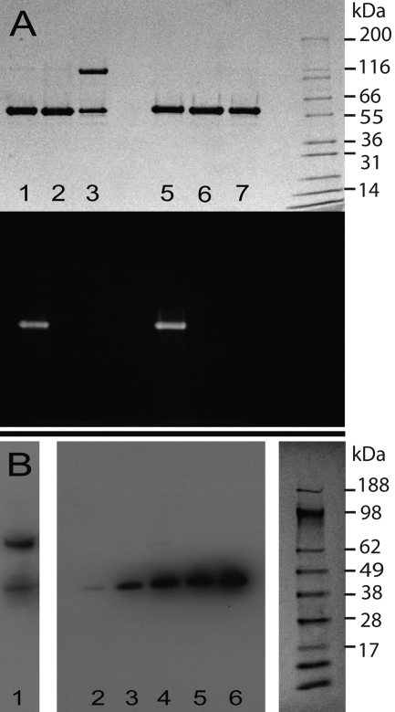
FIGURE 2.
Secretion of SkzL by S. agalactiae. A, protein-stained (top) and fluorescence (bottom) SDS-PAGE of nonreduced (lanes 1–3) and reduced (lanes 5–7) recombinant SkzL. [5F]-SkzL monomer (lanes 1 and 5), purified SkzL monomer (lanes 2 and 6), a 1:1 mixture of SkzL monomer and SkzL dimer (lanes 3 and 7), with the migration positions of molecular mass markers indicated (kDa). B, Western blotting of a mixture of recombinant SkzL monomer and dimer (lane 1) and proteins secreted from the lag, mid-logarithmic, late-logarithmic, early stationary, and late stationary growth phases (lanes 2–6), with molecular mass markers (kDa) from the corresponding Ponceau S-stained membrane. SDS-PAGE and blotting were performed, and membranes were probed with a polyclonal anti-SkzL antibody as described under “Experimental Procedures.”

