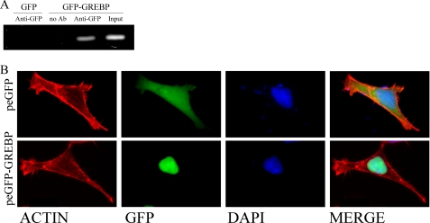
FIGURE 4.
A, ChIP assays for GREBP·cGMP-RE complex. HEK293 cells were transfected with peGFP or peGFP-GREBP for 12 h and fixed with formaldehyde as described under “Experimental Procedures.” GFP-overexpressing cells were used as negative controls. Fixed DNA·protein complexes were immunoprecipitated with an anti-GFP antibody, and normal rabbit IgG served as (no antibody) negative control. Input represented one-tenth of cleared unprecipitated supernatant. B, cellular localization of GREBP in HEK239 cells. HEK293 cells were transfected with peGFP or peGFP-GREBP for 12 h. They were then fixed and stained with phalloidin and 4′,6-diamidino-2-phenylindole. The upper panel shows the whole cell distribution of GFP as control. The lower panels present nuclear localization of the chimeric protein GFP-GREBP. DAPI, 4′,6-diamidino-2-phenylindole.

