FIGURE 3.
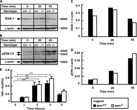
Stimulation of IRAK-1, ERK, and TNFα mRNA in asm+/+ and asm−/− macrophages. Peritoneal macrophages from asm+/+ and asm−/− mice were treated with LPS (10 ng/ml) for the indicated times. A–D, activation of IRAK-1 (A and C) and ERK1/2 (B and D). Analyses were done using Western blotting and antibodies against IRAK-1 and phosphorylated ERK1/2. Data are representative of three independent experiments. β-Actin levels were used to control for uniform loading. E, stimulation of TNFα mRNA. mRNA levels were determined by real time PCR. A glyceraldehyde-3-phosphate dehydrogenase (GAPDH) mRNA level was used for normalization. Data are presented as mean ± S.E. of three independent experiments. Statistical significance of the treatment effect was calculated (***, p < 0.001) based on two-way ANOVA.

