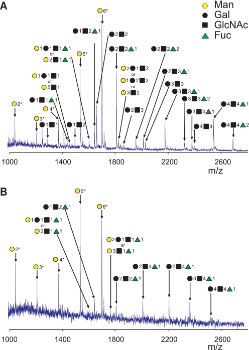
FIGURE 4.
Examples of N-glycan profiles from MALDI-TOF mass spectra. N-Linked oligosaccharides were isolated after digestion of gp120 expressed by HepG2 (A) or Jurkat cell lines (B) with PNGase F. Symbols with numbers indicate the number of saccharides added to the common core (GlcNAc)2(Man)3. Samples were prepared with PNGase F enzyme isolated from Flavobacterium that contained trace amounts of endo- and exoglycosidases that resulted in removing the reducing-end GlcNAc from high-mannose glycans (see “Experimental Procedures” for details). Identities of these glycans were confirmed by tandem mass spectrometry with LTQ mass spectrometry. *, denotes high-mannose glycans with (GlcNAc)1(Man)3 core. Representative results from two experiments are shown. Supplemental Fig. S3 shows MALDI-TOF mass spectra of glycans of the five gp120 preparations with molecular masses and compositions of detected glycans indicated.

