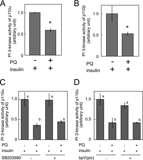
FIGURE 8.
Effect of paraquat on insulin-dependent activation of PI 3-kinase p110α and p110β. 3T3-L1 adipocytes were serum-starved for 18 h, and then cells were treated with 0 or 10 mm paraquat for 3 h followed by incubation with 10 nm insulin for 5 min at which time cells were solubilized. PI 3-kinase activity in immunoprecipitates by anti-p110α antibody (A and D) or anti-p110β antibody (B) was measured. C, 3T3-L1 adipocytes were serum-starved for 18 h, and then cells were treated with 0 or 10 mm paraquat in the presence or absence of 10 mm SB203580 for 3 h followed by incubation with 10 nm insulin for 5 min. PI 3-kinase activity in immunoprecipitates by anti-p110α antibody was measured. D, 200 nm dipotassium bisperoxo(picolinato)oxovanadate (V) (bpV(pic)) was added in the in vitro PI 3-kinase reaction mixture. The experiments were performed in triplicate, and the results shown are the mean ± S.E. * indicates the difference between cells with and without paraquat treatment is significant with p < 0.05 in A and B. There are significant differences between values with different superscript characters (p < 0.05) in C and D.

