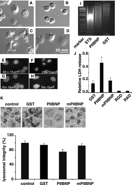
FIGURE 7.
PIIBNP induces death of tumor cell lines through a necrosis pathway. hCh-1 cells were plated on coverslips in a 6-well plate previously coated with type I collagen. The cells were incubated with 4 μm GST (A) for 60 h, staurosporine (STS) (B) for 16 h, PIIBNP (C) for 60 h, or mPIIBNP (D) for 60 h. The cells were imaged by a differential interference contrast light microscopy using a Q Capture Retiga 2000R camera (×40 magnification). The cell membrane integrity is lost in PIIBNP-treated cells (arrow, white) but not in staurosporine-treated cells (arrowhead, black). Cell nuclei from cells treated with GST (E), staurosporine (F), PIIBNP (G), and mPIIBNP (H) were stained with Hoechst and photographed by light microscopy using a Q Capture Retiga 2000R camera (×60 magnification). The area of the nucleus was measured using Northern Eclipse software. Nuclear area values in the panels are represented as mean ± S.D. (n = 30). The DNA laddering assays (I) indicated that the DNA integrity was lost only in cells treated with staurosporine (16 h) and not in cells treated with GST, PIIBNP, or mPIIBNP (90 h). Lactate dehydrogenase release assays (J) indicated that the relative LDH release occurred primarily in cells treated with PIIBNP (10 μm, 90 h) and much less in cells treated with GST (10 μm, 90 h), mPIIBNP (10 μm, 90 h), RGD (1 mm, 90 h), or RAD (1 mm, 90 h) synthetic peptides. K, neutral red lysosomal retention assay. The top panel shows neutral red retained in hCh-1 cells. Neutral red retention was quantified by absorption at 540 nm after extraction with 0.5 m HCl, 50% ethanol. The percentage of lysosomal integrity was calculated from the absorption values.

