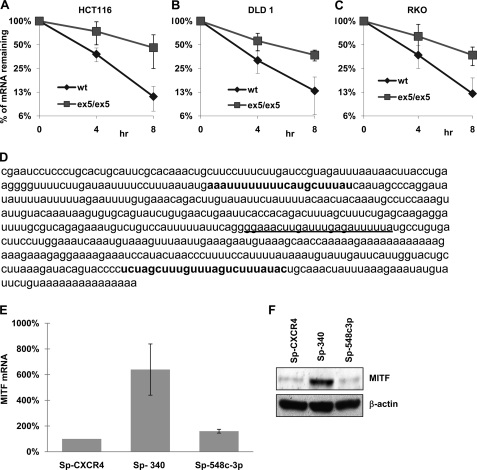
FIGURE 2.
The abundant MITF mRNA with short 3′-UTR is also regulated by miRNA. A, Dicerwt and DicerEx5/Ex5 HCT116 cells were co-transfected with Tet-off plasmid and p-BIG-MITF plasmid with short 3′-UTR. Transcription was stopped by treatment with doxycycline for the indicated durations. The stability of MITF transcripts was analyzed by measuring MITF mRNA levels with real time RT-PCR (normalized to GAPDH expression). B, p-BIG-MITF plasmid with short 3′-UTR was expressed in DLD1 cells, Dicerwt (wt), and DicerEx5/Ex5 (ex5/ex5) under the control of the Tet-off system. The stability of MITF mRNA was analyzed as in A. C, stability of MITF transcript with short 3′-UTR expressed in RKOwt and RKO DicerEx5/Ex5 cells was analyzed as in A. All of the results are representative of three separate experiments and are expressed as the mean values ± S.D. (error bars). The average half-lives of mRNAs are presented in supplemental Table S3. D, sequence of the short 3′-UTR of MITF showing binding sites for miR-340 (in bold type) and miR-548c-3p (underlined). E, the levels of endogenous MITF mRNA in 451Lu cells, transfected with the indicated miR-Sponge constructs, were estimated by real time RT-PCR after normalization with respect to GAPDH expression. The results are representative of three separate experiments and are expressed as percentages of control (SP-CXCR4) as the mean values ± S.D. (error bars). F, immunoblot analysis of MITF expression in the 451Lu cells transfected with the indicated miR-Sponge constructs (upper panel). The lower panel shows the expression of β-actin. The levels of endogenous miR-340 are presented in supplemental Fig. S1A.

