FIGURE 7.
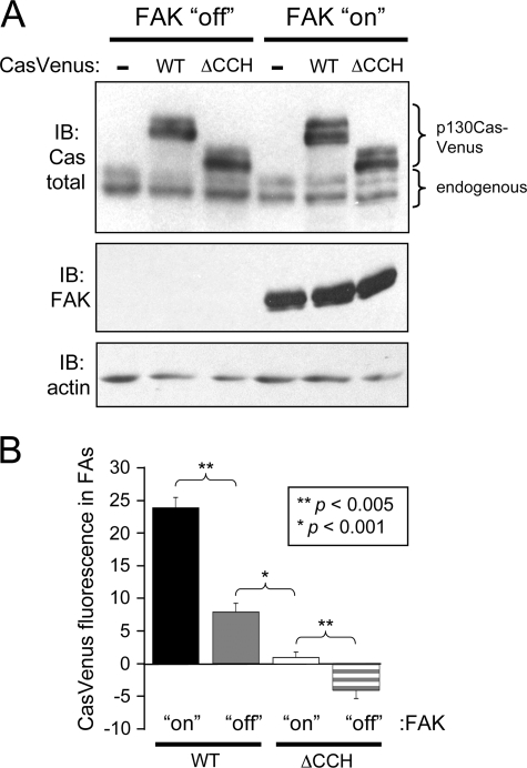
FAK plays a role in localizing p130Cas to FAs. A, shown is an immunoblot (IB) analysis of whole cell lysates from TetFAK cells expressing either p130CasVenus(WT) or p130CasVenus(ΔCCH) that were either induced to express FAK or kept in the non-induced condition. Detection with a total p130Cas antibody (top) shows the expression of the two p130CasVenus variants in comparison to endogenous p130Cas. A total FAK antibody was used to confirm the induced FAK expression (middle), whereas actin detection was used as a loading control (bottom). B, quantitative assessment is shown of the localization of WT and ΔCCH p130CasVenus variants to FAs in the presence or absence of FAK expression. Paxillin immunofluorescence was used to delineate FA borders, and mean Venus-YFP fluorescence values were measured from within the FA boundary. For each p130Cas variant, mean Venus-YFP fluorescence was determined from 50 FAs (10 each from 5 separate cells). Bars indicate S.E., and statistical significance was determined by Student's t test.

