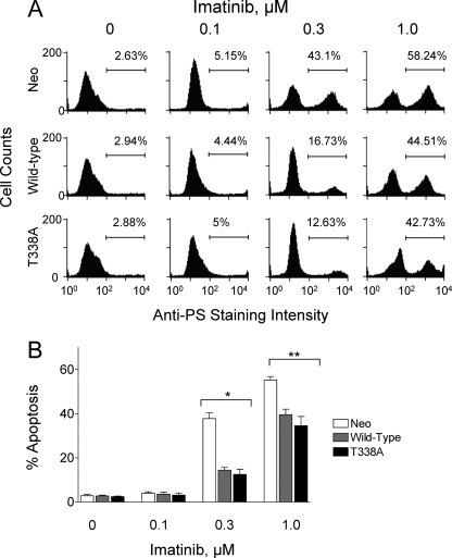
FIGURE 3.
Expression of wild-type Hck or Hck-T338A protects K562 cells from imatinib-induced apoptosis. K562-Neo, K562-Hck, and K562-Hck-T338A cell populations were incubated for 72 h in the absence or presence of the indicated concentrations of imatinib. Apoptotic cells were stained with an anti-phosphatidylserine-Alexa Fluor 488-conjugated antibody and the percentage of apoptotic cells was determined by flow cytometry. A, shown are histograms from a representative flow cytometry experiment with the percentage of apoptotic cells shown above each plot. PS, phosphatidylserine. B, bar graph showing the average percentage of apoptotic cells from three independent experiments ±S.D. Two-way ANOVA showed significant effects of Hck or Hck-T338A expression (p < 0.0001) and of imatinib treatment (p < 0.0001). One-way ANOVA was performed separately for each concentration of imatinib across the three groups. The percent apoptosis among the three cell lines was statistically significant at 0.3 μm imatinib and 1 μm imatinib (*, p = 0.0003 and **, p = 0.006). Bonferroni's post test for multiple comparisons showed a p < 0.001 for Neo versus Hck and Neo versus Hck-T338A at 0.3 μm imatinib and a p < 0.05 for Neo versus Hck and Neo versus Hck-T338A at 1 μm imatinib.

