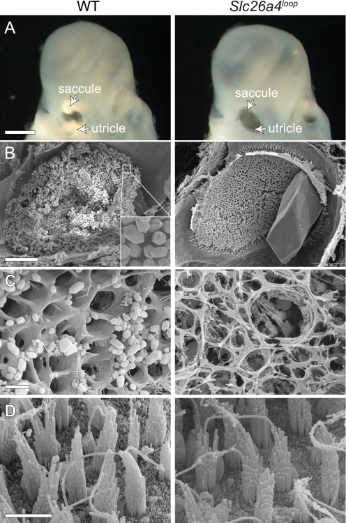
FIGURE 3.
Gross malformation of the vestibular gravity receptor. A, wild-type otoconia can be easily detected under a light microscope when looking at isolated inner ears. Two bright reflected areas represent the otoconia of the saccule and utricle of newborn mice. Slc26a4loop mutant mice lack the bright reflection, and instead, a dark hole appears when looking through the oval window (arrowheads). B, SEM of 2-month-old utricle shows thousands of otoconia (inset) that cover the sensory epithelium in a wild-type mouse, whereas a giant stone is appearing at Slc26a4loop/loop utricle. C, imaging of the gelatinous matrix (otoconial membrane) reveals its normal structure in wild-type mice, which is characterized by typical pores. In Slc26a4loop mutants, the otoconial membrane is dissociated and severed. D, high resolution images of the underlying hair cells show that Slc26a4loop/loop vestibular hair cells appear to be normal. Scale bars equal 500 μm in panel A, 100 μm in B, 5 μm in C and D. n = 4.

