FIGURE 6.
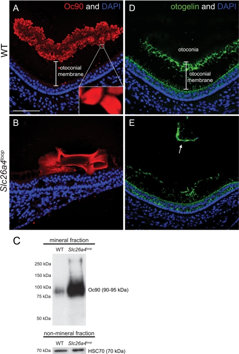
Oc90 conglomerates assemble the core of the calcitic giant minerals. A and B, Oc90 immunostaining on P15 paraffin sections of decalcified inner ears. A, wild-type otoconia are labeled with Oc90 (red) and highlight the boundaries of each otoconium. The unlabeled gap between the sensory epithelium (nuclei) and the otoconia represents the gelatinous matrix (otoconial membrane) that supports the otoconia load. B, a Oc90 (red) conglomerate is present in the giant calcitic mineral. Unlike the smooth expression of Oc90 in a single otoconium (inset in A), in the giant mineral, Oc90 expression has different levels of intensity along the mineral body. Furthermore, the giant mineral resides directly on the sensory epithelium, and the gap that corresponds to the otoconial membrane is absent. C, Western blot analysis of Oc90 in the mineral fraction shows a dramatic increase of protein content in the giant minerals. As a control, the non-mineral fraction was loaded on a gel and labeled with HSC70, confirming the equal amount of pooled utricles that were analyzed in each sample. D and E, otogelin immunostaining on P15 paraffin sections of decalcified inner ears. D, in wild-type mice, otogelin (green) is expressed by the supporting cells of the sensory epithelium. The secreted otogelin participates in the otoconial membrane assembly and maintenance. E, in Slc26a4loop mutant mice, otogelin is expressed by supporting cells; however, the otoconial membrane is severed, disorganized and displaced (arrow in D). Scale bars equal 75 μm in panels A–D. n = 5.

