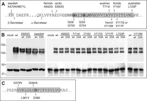
FIGURE 1.
APP- and PS1-FAD mutations. A, part of the APP sequence is shown. Indicated are the β- and α-secretase cleavage sites as well as all analyzed APP-FAD mutations. The glycine residues mediating the APP-TMS dimerization (9) are highlighted (Gly29/Gly700 and Gly33/Gly704 according to Aβ or APP770 numbering, respectively). B, APP-FAD and APP-FAD-G33A constructs are equally well and stably expressed in the SH-SY5Y cells. The double band represents mature, plasma membrane-residing (∼130 kDa) and immature (∼110 kDa) APP, and Western blot was stained with 22C11. As loading control, actin was stained as shown in the lower panel (∼40 kDa). C, an amino acid sequence of PS1 TMS-7 is shown. Analyzed FAD mutations and the catalytically active Asp385 are highlighted. The gray boxes in C mark residues embedded in the cell membrane.

