FIGURE 5.
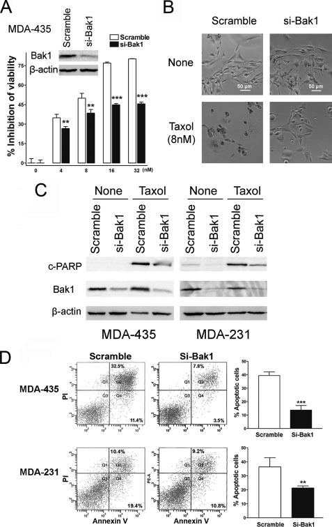
Bak1 plays a critical role in Taxol-induced apoptosis. A, MDA-435 cells were transfected with scramble siRNA or Bak1 siRNA (si-Bak1). Forty-eight hours after transfection, lysates were prepared, and Western blotting was carried out with antibody against Bak1 (inset). β-Actin was used as a loading control. MDA-435 cells with knockdown of Bak1 were seeded into 96-well plate at the density of 8 × 10³ per well and then followed with 0, 4, 8, 16, and 32 nm Taxol treatment for 48 h. Scramble siRNA served as a negative control. The inhibition of cell viability was detected. Columns, mean of three independent experiments; bars, S.E. *, p < 0.05, **, p < 0.01, ***, p < 0.001. B, MDA-435 cells with Bak1 knockdown by specific siRNA to Bak1 were seeded into 96-well plates at 8 × 10³ cells per well. After 12 h incubation cells were treated with 8 nm Taxol for 48 h. The cells were visualized using a phase-contrast microscope. Scramble siRNA-transfected MDA-435 cells served as a negative control. C, MDA-435 and MDA-231 cells were transfected with 100 nm Scramble siRNA (Ctr) or si-Bak1, 24 h after transfection, and cells were treated with 20 lysates were prepared for Western blotting with the antibodies against cleaved PARP (top) and Bak1 (middle). β-Actin was used as a loading control (bottom). D, MDA-435 and MDA-231 cells were transfected with 100 nm Scramble siRNA (Ctr) or si-Bak1, and 24 h after transfection cells were treated with 20 or 40 nm Taxol for 48 h, respectively. Then the cells were collected for apoptosis analysis by annexin V staining and flow cytometry. The percentage of apoptotic cells are represented in bar diagram from three independent experiments.

