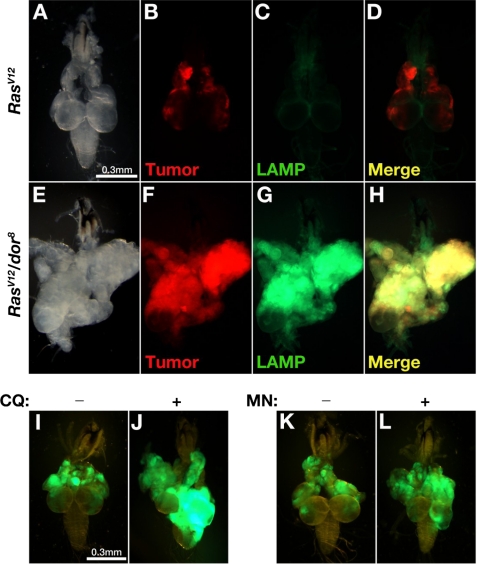
FIGURE 4.
Impaired lysosomal degradation contributes to metastasis. Dissected cephalic complexes from third-instar larvae are shown. A–H, RFP was used for labeling tumor clones to distinguish them from the green signal of the GFP-LAMP fusion protein. I–L, RasV12 tumor cells were labeled by GFP. In RasV12 tumors (A–D), GFP-LAMP fusion proteins were presumably targeted to lysosomes and efficiently degraded. With additional dor8 mutation, the lysosomal degradation of GFP-LAMP proteins was blocked inside tumor tissues with invasive potential (E–H). Inhibition of lysosomal degradation by feeding chloroquine (CQ) (J) or monensin (MN) (L) induced VNC invasion of RasV12 tumor clones (green) when compared with the control samples (I and K).

