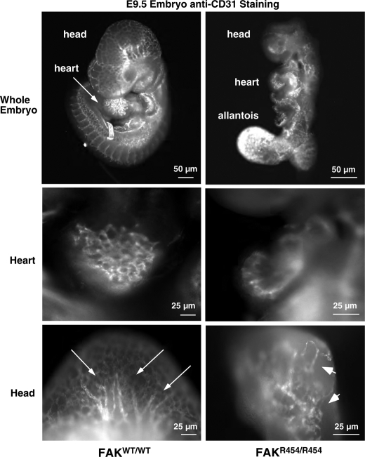
FIGURE 2.
Defective EC patterning within FAKR454/R454 embryos. Whole embryos at E9.5 were stained with anti-CD31 to visualize ECs. Comparisons are between FAKWT/WT and FAKR454/R454 littermates. Defined EC branchlike and tubule structures (arrows) in the head and heart of FAKWT/WT embryos are shown. FAKR454/R454 embryos exhibit intense EC staining in the unfused allantois. Clusters (arrowheads) of ECs are present in FAKR454/R454 head and heart structures, but these were not organized within a defined network.

