
FIGURE 3.
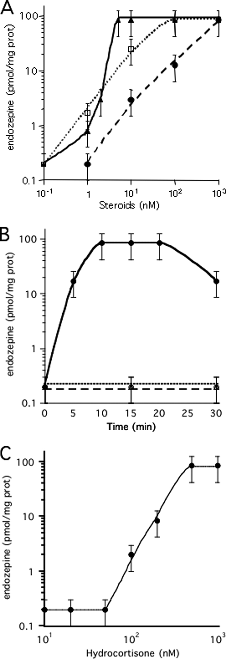
Induction of endozepine production by steroids. A, induction by pregnenolone sulfate and cortisol in the presence or absence of mifepristone. The indicated concentrations of cortisol (filled triangles) or pregnenolone sulfate (open circle) were added to astrocytes. For competition assay, 1 μm mifepristone was added to astrocytes just prior to endozepine induction with the indicated amount of cortisol (open circle) or pregnenolone sulfate (filled circle). The cells were incubated 15 min at 37 °C before harvesting 400 μl of the buffer to quantify the amount of endozepines with the Dictyostelium bioassay. The experiments were repeated at least three times. The bars indicate the reliability range. B, time course of endozepine production. Endozepine production was induced by addition of 100 nm cortisol to the astrocytes (circles) incubated at 37 °C for the indicated time. 2 μm TPCK (squares) or anti-ACBP antibodies (triangles) diluted 1/5,000 were added just prior to induction by cortisol. The amount of endozepine produced in a well was quantified. The experiments were repeated at least three times. The bars indicate the reliability range. C, induction by pregnenolone and progesterone. The indicated concentrations of pregnenolone (filled triangles) or progesterone (open square) were added to astrocytes. The amount of endozepine was quantified from samples harvested after 15 min incubation at 37 °C. The experiments were repeated at least three times. The bars indicate the reliability range.