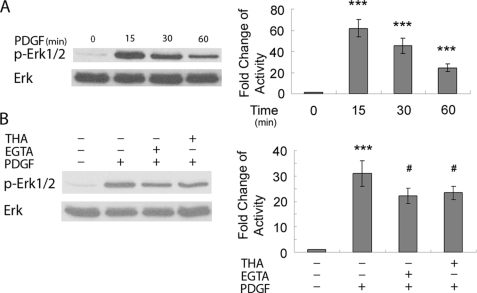
FIGURE 5.
PDGF-induced calcium transients lie upstream of MAPK/ERK pathway. A, exposure of rat hippocampal neurons to PDGF lead to time-dependent increase of ERK activation is shown. ***, p < 0.001 versus control group. B, pretreatment of rat hippocampal neurons with either EGTA or thapsigargin resulted in partially deactivation of ERK compared with PDGF treatment alone. ***, p < 0.001 versus control group; #, p < 0.05 versus PDGF (20 ng/ml) group. A representative immunoblot and the densitometric analysis from four separate experiments is presented. THA, thapsigargin.

