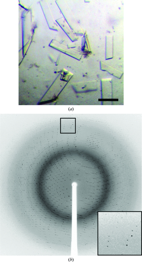
Figure 3.
Crystals and a sample diffraction image from PPM cystallized with Mn2+. (a) Crystals of PPM grown in the presence of 50 mM MnCl2. Optimized crystals of PPM that were identifed after supplementing the protein with 50 mM MnCl2. Crystals grew from 100 mM bis-tris pH 5.5, 50 mM MnCl2, 13–16% PEG 3350 and 25–100 mM ammonium acetate and appeared in 3–5 d at 291 K. The black bar represents 100 µm. (b) Diffraction from crystals of PPM grown in the presence of 50 mM MnCl2. Diffraction images collected at APS on beamline ID-21-G using 1° oscillations per frame revealed that these crystals belonged to space group P21. A data set consisting of 180 frames and is 98.6% complete to 1.85 Å was collected.

