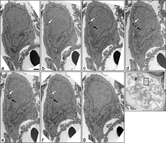
Fig. 3.

Serial transmission electron micrographs of a mature podocyte in adult rat (a–g). These sections include both the mother (solid arrows) and daughter (open arrows) centrioles, which are located near the nucleus. The mother centriole is not in touch with the surface plasma membrane, and no primary cilium was projected from the mother centriole. Bar 1 µm. h The podocyte shown by the serial sections is located in the rectangle
