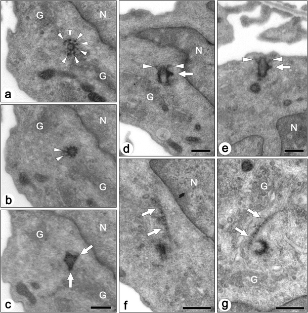
Fig. 4.

Transmission electron micrographs of centrioles and associated structures in the mature podocytes of adult rats. Axial (a–c) and longitudinal (d,e) sections of mother centrioles show transitional fibers (arrowheads) and basal feet (arrows). Even in the case where the mother centriole is close to the surface plasma membrane, the transitional fibers are not in contact with the membrane as indicated by arrowheads in (e). a–c Axial serial sections. f,g Striated rootlets (arrows) are occasionally found near the centrioles. G Golgi apparatus, N nucleus. Bars 500 nm
