
Fig. 8.
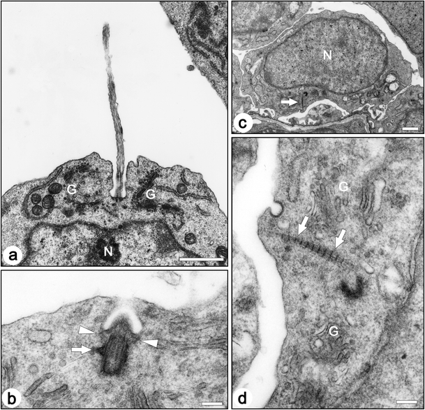
Transmission electron micrographs of centrioles and associated structures in the immature podocytes of fetal rats. a In most cases, a primary cilium protruded from the bottom of the membrane recess. b A longitudinal section of a basal body/mother centriole shows transitional fibers (arrowheads) and a basal foot (arrow). In ciliated podocytes, the basal body/mother centriole is located beneath the bottom of the membrane recess, and the transitional fibers are in contact with the inner side of the bottom membrane. c,d The striated rootlets (arrows) are frequently found near centrioles. G Golgi apparatus, N nucleus. Bar 1 µm (a, c), 200 nm (b,d)