Fig. 2.
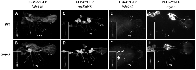
cwp-5(tm1893) does not disrupt RnB cilium structure or PKD-2 localization. Images show the localization of the indicated green fluorescent protein (GFP) fusion protein in wild-type (A,C,E,G) and cwp-5(tm1893) (B,D,F,H) adult male tails. In all panels, arrowheads indicate the cilium of R3B (the RnB neuron of ray 3). Insets show high-magnification views of the R3B cilia. Bars, 10 μm (A,B). All panels show lateral views, with anterior to the left, except for the ventral views in E,F.

