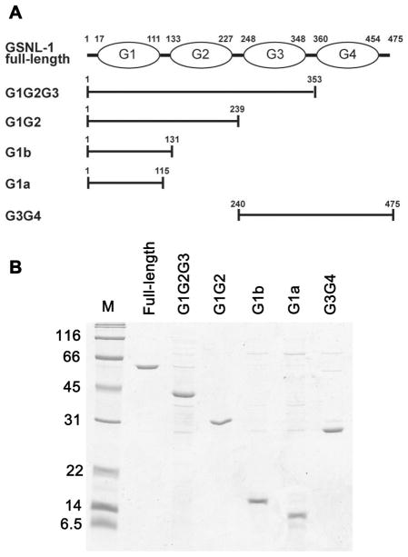
Figure 1.
Expression and purification of GSNL-1 variants. (A) Schematic representation of domain structure of full-length GSNL-1 (top). G domains are designated as G1 to G4. Positions of N- and C-termini of each domain are shown on top. Positions of amino acid sequence for each GSNL-1 fragments are shown. (B) Purified recombinant GSNL-1 variants (0.5 μg each) were subjected to SDS-PAGE and stained by Coomassie Brilliant Blue. Molecular mass markers (M) in kDa are shown on the left of the gel.

