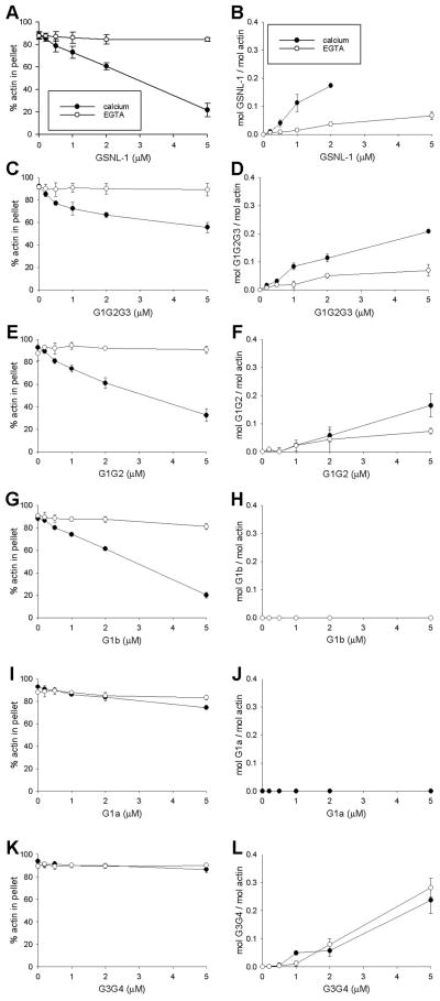
Figure 6.
Co-sedimentation assays of GSNL-1 variants with F-actin. 5 μM F-actin was incubated with varying concentrations (0 – 5 μM) of full-length GSNL-1 (A and B), G1G2G3 (C and D), G1G2 (E and F), G1b (G and H), G1a (I and J), or G3G4 (K and L) in the presence of 0.1 mM CaCl2 (closed circles) or 0.1 mM EGTA (open circles) for 1 hr and ultracentrifuged at 436,000 × g for 15 min. The supernatants and pellets were separated and analyzed by SDS-PAGE. Band intensities were quantified by densitometry, and actin-independent sedimentations of GSNL-1 variants were subtracted from the experimental data in the presence of actin. On the left column (A, C, E, G, I, and K), percentages of actin in the pellet fractions are plotted as a function of total concentrations of GSNL-1 variants. On the right column (B, D, F, H, J, and L), molar ratios of GSNL-1 variants to actin in the pellets are plotted as a function of total concentrations of GSNL-1 variants. Data are average ± standard deviations of three experiments.

