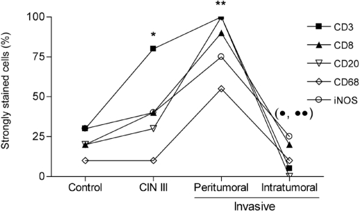
Figure 2.
Distribution of immunohistochemically labeled cells. T lymphocytes (CD3+ and CD8+), B lymphocytes (CD20+), macrophages (CD68+) and cells that express iNOS from patients in the control group, the CIN III group and the group with invasive carcinomas of the uterine cervix in the peritumoral stroma and the intratumoral microenvironment were detected by immunohistochemistry as described in the “Materials and Methods” section. * P < 0.002, CD3+ compared with control; ** P < 0.01, all markers compared with CIN III (except CD3 and iNOS) and control; • P < 0.005, all markers compared with peritumoral; •• P < 0.05, CD3+ and CD20+ compared with CIN III.

