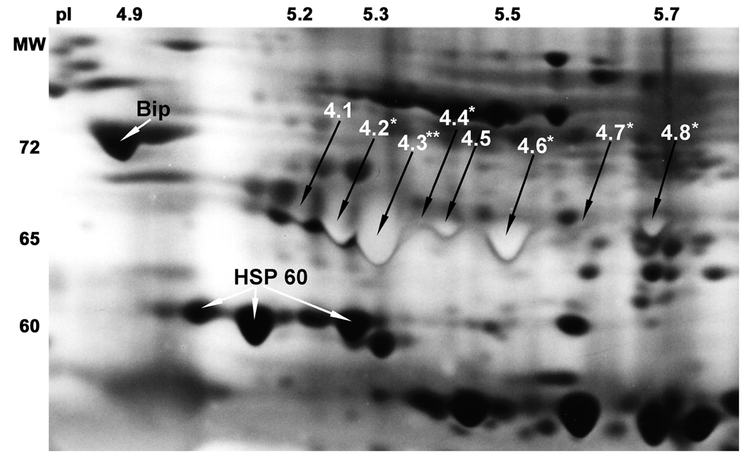
Figure 2.
Expansion of the sperm proteome in the region showing the position of the microsequenced 65 kDa HSP70 proteins.
Enhanced resolution of region of interest was achieved with an ampholine composition of 20% pH 3.5–5, 25% pH 5–7, 10% pH 7–9 and 45% pH 3.5–10 in the first dimension. The unique light staining of the eight 65 kDa surface exposed HSP70 forms was achieved by using four times the normal concentration of ammonium hydroxide in the silver nitrate solution, aiding their identification and excision from silver stained gels. The eight proteins in position 4 are numbered from 4.1 to 4.8. Amino acid substitutions were found in peptides from 6 of the protein spots (stars). The positions of Bip (HSPA5) and HSP60 (HSPD1) are indicated by white arrows. The most acidic HSP60 form was microsequenced, while the two basic forms were identified by immunoblotting.

