Figure 3.
Immunogenecity of human sperm HSP
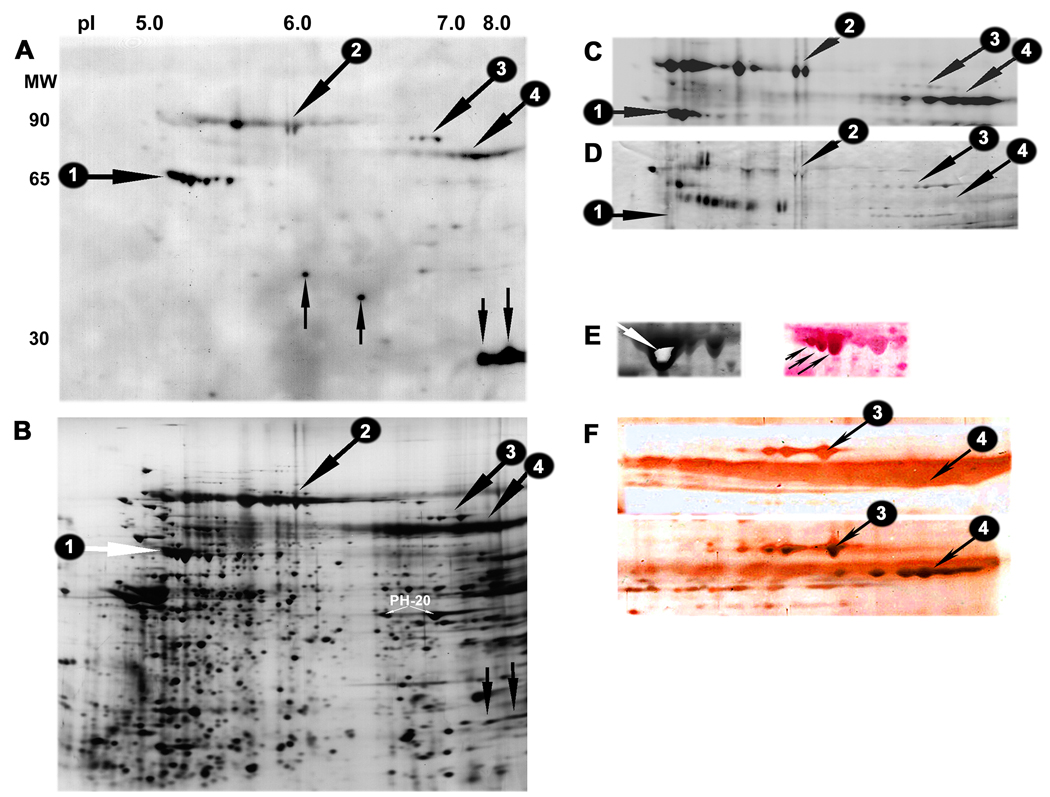
A: Two dimensional western blot demonstrating anti-sperm IgG antibody reactivity in patient serum number 629 towards seven of the eight HSP70 forms detected on the surface of human sperm (group #1 indicated by a horizontal arrow), as well as against two slightly basic proteins migrating at MW 27 kDa (downward vertical arrows). The serum also reacted with two sperm antigens of MW 34 kDa and 37 kDa, and three groups of proteins with MW 90–93 kDa, 84 kDa and 78–79 kDa, (respectively numbered 2–4 and indicated by oblique downward arrows).
B: Companion silver stained IEF/PAGE gel showing the position of the major antigens or groups of antigens detected by serum 629 in blot A.
C: Biotin label demonstrating surface exposure of the high molecular weight sperm antigen groups 1–4. The remaining major sperm antigens recognized by the serum were not accessible for surface labelling with neither biotin nor radioiodine (data not shown).
D: Two dimensional affinity blotting with HRP-conjugated Concanavalin A (ConA) demonstrating relatively strong staining of group 3 human sperm surface antigens. The HSP70 surface antigens did not bind ConA, while weak staining of the antigens in group 2 & 4 was observed.
E: Coomassie stained HSP70 forms from large preparative gel (left). The center of the most abundant HSPA2 isoform (spot 4.3 in Figure 2) was excised (oblique white arrow) and used as immunogen in female rabbits. The right image shows the same gel area on a gold stained NC-membrane. The membrane was subsequently stained with the antiserum raised against the excised HSPA2 form. Note the immunostaining of several HSP70 isoforms with approximate MW 65 kDa, including the three strongly stained species indicated by oblique black arrows.
F: High MW area of gold stained membranes incubated with the preimmune rabbit serum (top) and the antiserum against testis-specific HSP70 (bottom). Note the strong staining of sperm surface antigen groups 3 and 4 with the HSP70 antiserum.

