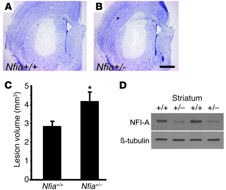
Figure 7. NFI-A–deficient neurons are sensitive to NMDA treatment in vivo.
(A and B) Representative photomicrograph of intrastriatal lesion of (A) Nfia+/+ mice and (B) Nfia+/– mice 2 days after microinjection of 0.3 ml NMDA (50 mM). Scale bar: 50 mm. (C) Quantification of lesion volumes. n = 4; *P < 0.01, Student’s t test. (D) Immunoblot of NFI-A expression in the striatum of Nfia+/+ and Nfia+/– mice.

