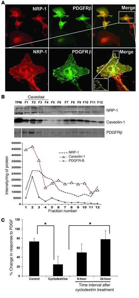
Figure 2. NRP-1 is enriched within low-buoyant-density membrane microdomains and colocalizes with PDGFRβ.
(A) LX2 cells transduced with PDGFRβ retrovirus were cotransduced with Ad-NRP-1–RFP. Cells were immunostained with NRP-1 (red) and PDGFRβ (green) Abs and photographed under a confocal microscope. The top row shows ×20 magnification and the bottom row shows ×63 magnification of the cropped cell, with further magnification of the cropped area (representative photomicrographs or Western blot are from 3 independent experiments). (B) HSC lysates were prepared for sucrose density gradient fractionation studies, and equal volumes of each fraction were analyzed by SDS-PAGE (top). White line indicates splicing of a noncontiguous sample from the same membrane. NRP-1 was also found to be enriched within low-buoyant-density fractions as assessed by levels of NRP-1 per microgram protein within each fraction when SDS-PAGE fraction signal was normalized for protein concentration (bottom). Separation and purity of membrane fractions were determined by immunoblotting for caveolin-1, a marker for low-buoyant-density membranes (n = 3). TPM, total plasma membrane. (C) Cyclodextrin, a compound that disrupts low-buoyant-density vesicle formation, inhibits PDGF-dependent hHSC chemotaxis. Cyclodextrin inhibition of PDGF-induced HSC chemokinesis was studied using time-lapse video microscopy followed by analysis using MetaMorph Imaging software. The right 2 bars show a reversible effect of cyclodextrin, thus reducing the likelihood of cell toxicity relating to the compound (n = 3, *P < 0.05).

