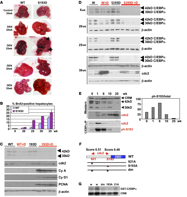
Figure 3. Development of tumors is accelerated in C/EBPα-S193D knockin mice.
(A) Pictures of livers at different time points after DEN injection. Arrows show large tumor nodules observed in livers of mice treated with DEN for 25 weeks. (B) DNA synthesis is increased in livers of S193D mice. Bar graphs show percentage of BrdU-positive hepatocytes in tumor sections of livers of WT and S193D mice. Summary of results with 3 animals of each genotype is shown. Data shown are mean ± SD. (C) Expression of C/EBPα and cell-cycle proteins in WT and S193D livers at 25 weeks after DEN injection. Nuclear extracts were examined by Western blotting with antibodies shown on the right. (D) The mutant S193D isoform of C/EBPα is specifically degraded during liver tumor development. Western blotting was performed with WT and S193D livers at 6, 20, 25, and 30 weeks after injection of DEN. 2 isoforms of C/EBPα are shown by arrows. (E) cdc2 is elevated during DEN-mediated carcinogenesis and phosphorylates C/EBPα at S193 before degradation of C/EBPα. Western blotting was performed with nuclear extracts of WT mice. Bottom: C/EBPα was IP from nuclear extracts, and the IPs were probed with antibodies to cdc2 and ph-S193 C/EBPα. Bar graphs show a ratio of S193-ph isoform of C/EBPα to total C/EBPα protein. (F) Location of the predicted cdc2 sites within C/EBPα and generation of mutant constructs. (G) Cdc2 phosphorylates C/EBPα at S193 and S21. Cdc2 was IP from nuclear extracts isolated from the liver at 30 weeks after DEN treatments. Kinase assay was performed with WT C/EBPα and with C/EBPα mutants S21A, S193A, and S21A–S193A (Dm).

