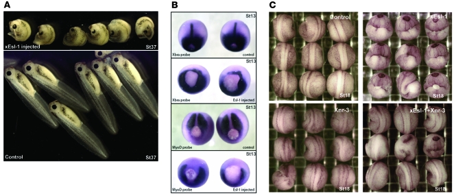
Figure 5. xEsl1 modulates Xenopus body axis formation.
(A) Morphological change of embryos (lateral view at stage 37) after injection at 2-cell stage. Upper panel: Embryos injected with xEsl1 mRNA (460 pg) in the dorsal marginal zone display shortened and curved body axis. Lower panel: Control embryos injected with xEsl1 mRNA (460 pg) in the ventral marginal zone show no change in phenotype. (B) The xEsl1 injection leads to changes of mesoderm markers detected by whole mount in situ hybridization (dorsal view at stage 13 [St13]). Top panel: Xbra was expressed around the blastopore and at notochord midline in the uninjected embryos. Second panel: Xbra expression was reduced or absent in xEsl1 mRNA–injected embryos. Third panel: MyoD was mainly expressed in presomatic plate in the uninjected embryos. Bottom panel: MyoD expression was absent in presomatic plate of xEsl1 mRNA–injected embryos. (C) Rescue of xEsl1-induced axis defects by coinjection with Xnr3 mRNA (dorsal view at stage 18). Embryos were injected at 2-cell stage in the dorsal marginal zone with the indicated mRNAs, xEsl1 (300 pg), Xnr3 (13 pg), or both. The coinjection of Xnr3 mRNAs rescued the curved body axis and open neural fold phenotypes of xEsl1 overexpression.

