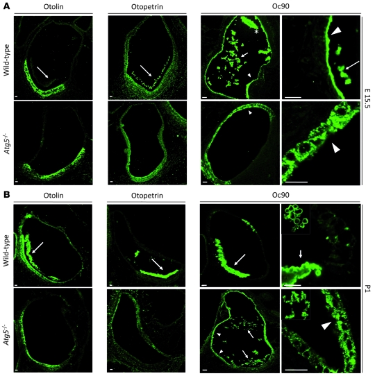
Figure 6. The correct localization of otoconial core proteins is dependent on autophagic activity.
(A) Representative immunofluorescence images of endogenous otolin, otopetrin, and Oc90 proteins in utricles from WT and Atg5–/– E15.5 embryos showing their mislocalization in the absence of autophagy. Both otolin and otopetrin presented a similar intracellular localization both in WT and Atg5–/– samples but were only detected in utricle lumen in autophagy-competent embryos (arrows). Oc90 (right) was mainly detected in WT samples inside utricle lumen, both at the developing otoconia (asterisk) and as a proteinaceous coral-like structure (arrows). In addition, Oc90 was also detected covering utricle epithelial and sensory cells in WT samples (arrowheads in top panels). In contrast, in Atg5–/– samples Oc90 was only detected inside non-sensory vestibular cells (arrowheads in bottom panels). (B) In P1 WT neonates, otolin and otopetrin were mainly detected in otoconial cores (arrows), whereas this localization was not observed in Atg5–/– samples. Similarly, in WT samples Oc90 was mainly detected in otoconial cores (right panel and arrow in left panel). In autophagy-impaired neonates, Oc90 localized both in utricle lumen, forming proteinaceous coral-like structures (right panel and arrows in left panel), and inside non-sensory cells (arrowheads). Scale bars: 50 μm (otolin, otopetrin, and left Oc90 panels); 30 μm (right Oc90 panels).

