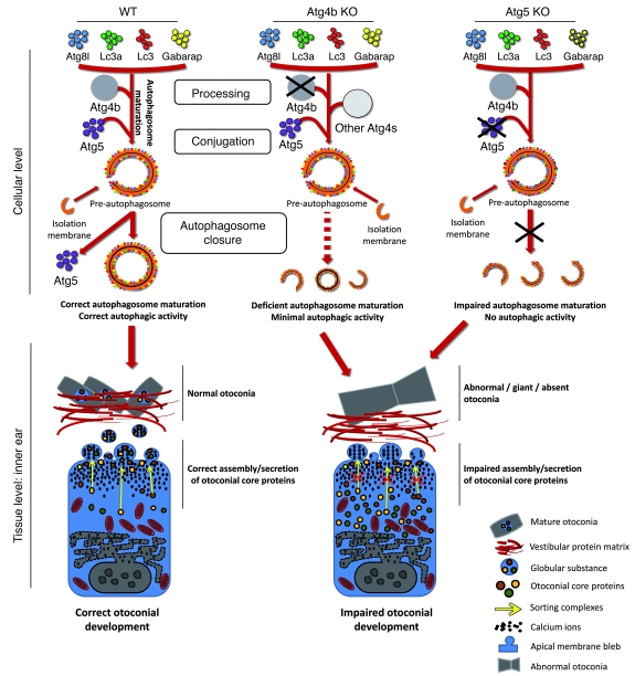
Figure 8. Proposed model for the otoconial defects observed in autophagy-impaired mice.
At the cellular level, Atg4b and Atg5 disruption affects autophagy by either reducing (Atg4b) or totally impairing (Atg5) autophagic activity. At the tissue level, these deficiencies lead to alterations in inner ear otoconial development. In fact, a severe reduction or a total disruption of autophagic flux in vestibular cells causes a dramatic impairment in the secretion and assembly of otoconial core proteins. This event is likely associated with transcriptional and other autophagy-dependent alterations of Ap-3 and Bloc-1 sorting complexes. All these cellular and tissue events lead to the development of abnormal or giant otoconia, or even to their total absence, finally resulting in the balance and coordination disorders observed in Atg4b–/– mice.

