
Fig. 3.
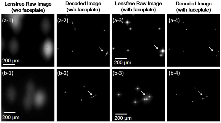
(a1) shows a digitally zoomed lensfree fluorescent image of 10µm particles that is obtained without the use of a faceplate. (a2) illustrates the output of the compressive decoder for the same image in (a1). (a3) shows the same region of interest imaged this time using a faceplate as shown in Fig. 1(a). The compressive decoder output of image (a3) is shown in (a4). The same story is repeated in (b1) through (b4) for a different region of interest. The arrows in these images specifically point to regions where the improvement due to the faceplate becomes apparent to better resolve closely spaced fluorescent particles.